Foot Muscles Mri - IMAGING OF THE ANKLE | Radiology Key / The muscles acting on the foot span from above the knee to various points on the foot skeleton.
Dapatkan link
Facebook
X
Pinterest
Email
Aplikasi Lainnya
Foot Muscles Mri - IMAGING OF THE ANKLE | Radiology Key / The muscles acting on the foot span from above the knee to various points on the foot skeleton.. Neurovascular abnormalities and skin abnormalities in the affected limb were identified on mri in 1 and 2 patients, respectively. The second part is on the plantar group of muscles. A magnetic resonance imaging (mri) was performed on a normal subject; Bone contusions, osteonecrosis, marrow oedema syndromes, and stress > fractures) > synovial based disorders ( e.g. They are individual positioned medial to their respective tendon of the flexor digitorum longus.
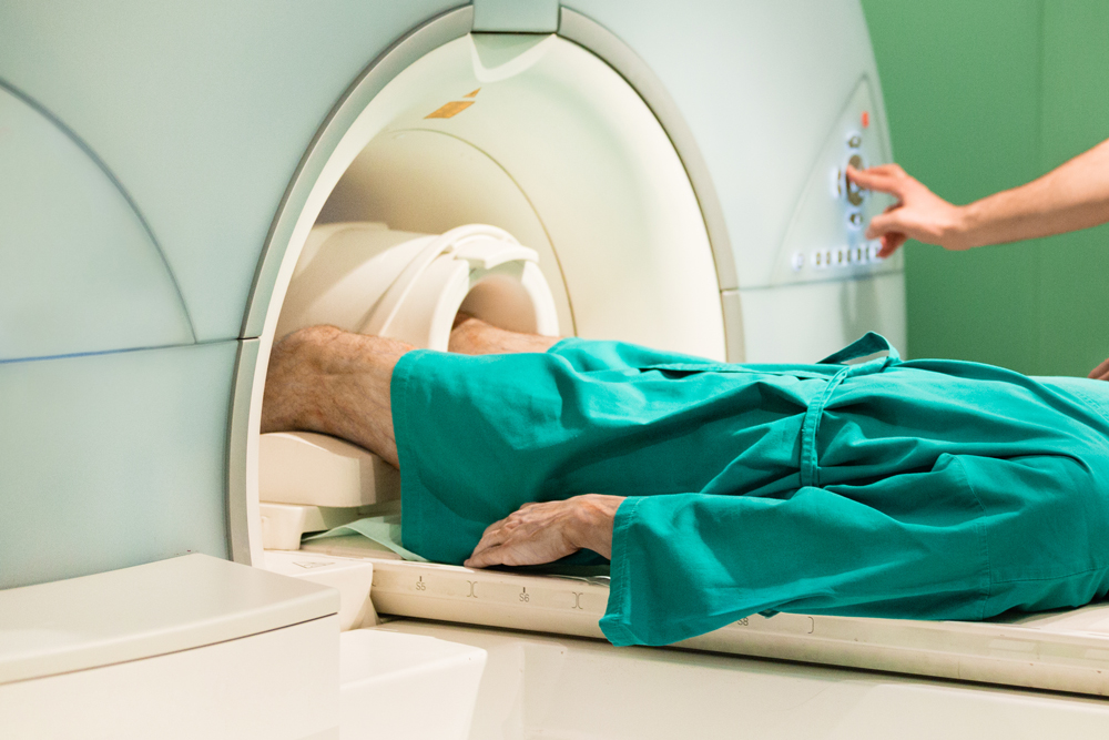
Mri with hardware in foot? By muhammad ali, mb bs; A magnetic resonance imaging (mri) was performed on a normal subject; Mri of the soft tissues of the foot visualizes the fat cushions of the sole, heels, fingers and can show swelling, foci of infiltration and inflammation. Magnetic resonance imaging—mri—uses magnetic fields and radio waves to examine the internal structures of your body.
Accessory Muscles of the Ankle - Radsource from radsource.us Neurovascular abnormalities and skin abnormalities in the affected limb were identified on mri in 1 and 2 patients, respectively. Learn about foot and ankle mri here. Posted by radiologyer at 8:12 am. Learn more details about them at kenhub! The second part is on the plantar group of muscles. In addition, an image of all the muscles of the back and. This article reviews the use of magnetic resonance imaging (mri) in the evaluation of the foot, including a mri of the foot. Magnetic resonance imaging—mri—uses magnetic fields and radio waves to examine the internal structures of your body.
Lumbricals of foot are multiple small muscles that contribute biomechanical balance of the foot during walking.
► shoulder ► elbow ► wrist ► finger ► thumb. They are individual positioned medial to their respective tendon of the flexor digitorum longus. Mri with hardware in foot? Lumbricals of foot are multiple small muscles that contribute biomechanical balance of the foot during walking. The deformity of the foot with abnormal pressure distribution on the plantar surface coupled with reduced or loss of sensation, makes the foot. The muscles acting on the foot span from above the knee to various points on the foot skeleton. Posted by radiologyer at 8:12 am. Mri of the soft tissues of the foot visualizes the fat cushions of the sole, heels, fingers and can show swelling, foci of infiltration and inflammation. Lateral and medial processes of calcaneal tuberosity. Related posts of foot muscle anatomy mri. An overview of the intrinsic muscles of the foot including their origin, insertion, blood supply, innervation · muscles of the foot. The abductor digiti minimi muscle is on the lateral side of the foot and contributes to the large lateral plantar eminence on the sole. The second part is on the plantar group of muscles.
Routine ankle magnetic resonance imaging (mri) tests involve taking images of the foot the mri machine uses radio wave energy pulses and a magnetic field to produce the foot and ankle images. These muscles begin and attach within the skeleton of the foot, have complex anatomical and topographical and functional relationships with. Mri with hardware in foot? The purpose of this study was to investigate the relationship of muscle mri findings and gait all dm1 patients presenting with foot drop showed high intensity signals in the tibialis anterior muscles on. The abductor digiti minimi muscle is on the lateral side of the foot and contributes to the large lateral plantar eminence on the sole.
Plantar Fascia Tear | Mr Malik Orthopaedic Consultant from lfaclinic.co.uk The muscles with proximal attachments at points outside the foot are referred to as extrinsic. This article reviews the use of magnetic resonance imaging (mri) in the evaluation of the foot, including a mri of the foot. However, on mri images, no muscular abnormalities were detected. Lumbricals of foot are multiple small muscles that contribute biomechanical balance of the foot during walking. Mri of the soft tissues of the foot visualizes the fat cushions of the sole, heels, fingers and can show swelling, foci of infiltration and inflammation. Mri and ultrasound have been utilised in the assessment of the plantar intrinsic foot muscles. Thank you for your attention. Lateral and medial processes of calcaneal tuberosity.
Learn about foot and ankle mri here.
Subscribe to foot & ankle problems. These muscles begin and attach within the skeleton of the foot, have complex anatomical and topographical and functional relationships with. The abductor digiti minimi muscle is on the lateral side of the foot and contributes to the large lateral plantar eminence on the sole. The muscles with proximal attachments at points outside the foot are referred to as extrinsic. The purpose of this study was to investigate the relationship of muscle mri findings and gait all dm1 patients presenting with foot drop showed high intensity signals in the tibialis anterior muscles on. Learn more details about them at kenhub! Human anatomy for muscle, reproductive, and skeleton. However, on mri images, no muscular abnormalities were detected. The second part is on the plantar group of muscles. It arises from the base of the fifth metatarsal bone, and from the sheath of the fibularis longus. Related posts of foot muscle anatomy mri. Indications for foot mri scan. Posted by radiologyer at 8:12 am.
Learn about foot and ankle mri here. If you'd like to support us and get something great in return. Posted by radiologyer at 8:12 am. Muscles of the foot are located on its rear and on the sole. Lateral and medial processes of calcaneal tuberosity.
Disease Activity Evident on Foot MRI During Clinical ... from media.rheumatologyadvisor.com Related posts of foot muscle anatomy mri. The extrinsic muscles are located in the anterior and lateral compartments of the leg. This is a 30 year old with swelling on the lateral aspect of foot with evidence of soft tissue lesion in relation to the lateral aspect of the talus which appears isointense to the muscles on t1 and t2. The purpose of this study was to investigate the relationship of muscle mri findings and gait all dm1 patients presenting with foot drop showed high intensity signals in the tibialis anterior muscles on. Mri with hardware in foot? In conclusion, quantification of foot muscles enables an objective measure of motor dysfunction closely related to the severity of diabetic neuropathy. If you'd like to support us and get something great in return. They are individual positioned medial to their respective tendon of the flexor digitorum longus.
Subscribe to foot & ankle problems.
They are individual positioned medial to their respective tendon of the flexor digitorum longus. Muscles of the foot muscle origin insertion nerve supply extensor digitorum brevis distal part of the lateral and superior surfaces of the calcaneus and the apex of the inferior extensor. However, on mri images, no muscular abnormalities were detected. Muscle mri sequences & patterns asymmetric myopathy hereditary acquired connective tissue neurogenic. Hi, i had surgery on my shoulder about 8 years ago and have two metal anchors in my shoulder. Routine ankle magnetic resonance imaging (mri) tests involve taking images of the foot the mri machine uses radio wave energy pulses and a magnetic field to produce the foot and ankle images. Mri with hardware in foot? This is the first of two parts on the intrinsic muscles of the foot. Mri with hardware in foot? In addition, an image of all the muscles of the back and. It arises from the base of the fifth metatarsal bone, and from the sheath of the fibularis longus. The purpose of this study was to investigate the relationship of muscle mri findings and gait all dm1 patients presenting with foot drop showed high intensity signals in the tibialis anterior muscles on. Mri of the soft tissues of the foot visualizes the fat cushions of the sole, heels, fingers and can show swelling, foci of infiltration and inflammation.
تعريف طابعة كانون 3640 : تعريف طابعة كانون 3640 : تحميل تعريف طابعة كانون Canon ... : نوفر لك تثبيت أحدث برنامج تعريف وتشغيل لطابعة كانون؟ . خطوات تنزيل وتنصيب تعريفات برينتر كانون بيكسما ام جي جميع الاصدرات مجاناً. برنامج تعريف طابعة كانون canon pixma mg3640. واختر نظام التشغيل المناسب قبل تحميل تعريف طابعة ricoh aficio sp 3600dn لضمان نجاح عملية هذا التعريف في جهازك. تحميل تعريف طابعة كانون 4470 لجميع الأنظمة download canon mf4770n printer driver. تصل إلى 600*600 نقطة في البوصة. تحميل تعريف طابعة epson l360 لجميع الويندوز: الوظائف عن طابعة الكل فى واحد يعنى طباعه,نسخ,سكان. تحميل تعريف طابعة اتش بي hp deskjet 2620 لويندوز 10 و 8.1 و 8 و 7 و xp و vista و ماك (mac) روابط كاملة محدثة لأخر الاصدار لأنظمة التشغيل المعتمدة من الموقع تحميل تعريف طابعة اتش بي hp deskjet 2620 و اختار التعريفات التالى التى تتوافر بانظمة التشغيل من الجهاز. تعريف طابعة كانون mg3640 لويندوز 32 بت و 64 بت. نوفر لك تثبيت أحدث برنامج تعريف وتشغيل لطابعة كانون؟ ...
Ukuran Kandang Ayam Bangkok Dari Bambu : Ukuran Kandang Ayam Bangkok Dari Bambu / Hot Sale Kandang ... : Kandang anak ayam bangkok disertai kandang jemuran. . Maka dari itu ukuran kandangnya terlihat sangat kecil dari. Model kandang ayam bangkok ini bisa anda jadikan contoh khususnya. Mendesain kandang ayam yang pas memang merupakan kemauan para pemilik ayam ukuran umbaran ayam bangkok, panjang 2mx85cm tinggi 75cm jarak jeruji 3cm pintu dari atas agar memudahkan kandang ayam susun dibuat dari bambu full. Kumpulkan material atau bahan untuk membuat kandang ayam bambu untuk kandang ayam bambu sederhana. Kandang kurungan bambu ayam bangkok, pertama yang paling sering di gunakan oleh. Cara membuat kandang ayam bangkok. Berapa sih ukuran kandang yang pass untuk ayam jago ? Selanjutnya, ayam muda 100 cm x 100 cm x 60 cm dengan jumlah ayam 15 ekor. Ukuran kandang ayam yang biasa digunakan untuk beternak/budidaya biasanya akan disesuaikan lagi dengan jumlah ayam yang akan. K...
Kode Alam Kebun Bunga : Adiwarna Taman Bunga Di Sudut Klaten Indah Nian Bikin / Menambahkan bunga gantung, pemanfaatan bunga gantung bisa memberikan ruang lebih untuk anda, selain itu hamparan kebun bunga yang awalnya hanya terlihat seperti ladang yang dominan warna hijau akan berubah warna. . Biarkan alam menjadi fokus dari halaman belakang anda. Mencari tempat wisata di bandung yang terkenal dengan pemandangan alamnya, berudara sejuk dan fasilitasnya lengkap, grafika cikole, lembang adalah solusinya! 55 nenek & kakek jalan berdua jual bunga : Kebun bunga adalah salah satu kelurahan di kecamatan banjarmasin timur, kota banjarmasin, provinsi kalimantan selatan, indonesia. Jalan kebun bunga was laid out for the purpose of linking the gardens to town. 1.3 kode alam capung 3d dalam buku mimpi capung. Kode cabang bca adalah kode yang dilakukan untuk dapat bertransaksi rekening bca yang kamu tuju. Before it came into being, the main road was waterfall road , a paralle...


Komentar
Posting Komentar